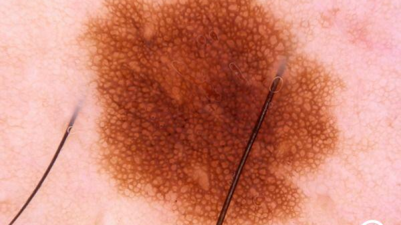
Moles, medically known as melanocytic nevi, are skin growths associated with pigment-producing cells.
They may be present at birth or develop later in life. Most moles are benign and do not require treatment.
However, some moles may change in shape, color, or size over time.
Mole monitoring aims to detect suspicious changes early while avoiding unnecessary removal of benign moles.
The widely recognized ABCDE rule can help assess moles:
Having one of these features does not necessarily mean a mole is cancerous, but it does warrant a dermatologic evaluation.
Skin lesions that look noticeably different from other moles, change rapidly, bleed without an obvious cause, or fail to heal should be examined promptly.
Dermoscopy is a painless examination that uses specialized lighting and magnification to assess moles and other skin lesions.
A device called a dermatoscope allows the dermatologist to examine pigment patterns, blood vessels, and structures within a lesion that are difficult to see with the naked eye.
This helps distinguish benign moles from suspicious pigmented lesions.
Dermoscopy does not involve needles or incisions.
Photographing moles at scheduled intervals and comparing the images can be helpful for some patients.
For people with numerous or atypical moles, digital dermoscopic monitoring and total-body photography may be performed using appropriate systems.
These methods can make subtle changes over time easier to identify.
However, not every mole or patient needs digital monitoring. A dermatologist determines the appropriate monitoring method and follow-up schedule.
A dermatologic evaluation is recommended if:
New pigmented lesions that appear and change during adulthood deserve careful assessment.
Not every mole needs to be removed.
Moles that appear benign on dermoscopy and cause no problems may be monitored.
However, a biopsy or surgical removal may be needed when a lesion raises concern for melanoma or another skin tumor.
In these cases, a definitive diagnosis is made through microscopic examination of the tissue.
No. Surgically removing a mole using an appropriate technique does not cause it to become cancerous.
What matters is assessing a suspicious mole correctly and sending tissue for pathologic examination when needed.
Pigmented lesions suspected of being melanoma should not be treated with laser or destroyed without pathologic examination.
Doing so can delay a definitive diagnosis.
There is no single follow-up interval that applies to everyone.
The schedule depends on the number and appearance of moles, a history of sunburn, a personal or family history of melanoma, and other risk factors.
Patients at higher risk may need closer monitoring.
Regularly checking your own skin and noticing new changes are also important.
Mole assessment aims to identify suspicious lesions promptly while avoiding unnecessary procedures.
At Dr. Armin Müştak’s private dermatology practice, moles are assessed through a dermatologic examination and dermoscopy.
Depending on the lesion’s features, the plan may include clinical monitoring, further evaluation, or surgical removal and histopathologic examination when appropriate.
Recognizing changes in moles early is important for the timely detection of skin cancer.
Moles that are asymmetrical, have irregular borders, contain multiple colors, grow, or change in appearance should be assessed. Bleeding without an obvious cause or a sore that does not heal are also warning signs. A dermatologic examination is needed to evaluate these changes.
No. Proper surgical removal of a mole does not cause cancer. However, suspicious moles should not be destroyed with laser or cautery without pathologic examination.
No. Dermoscopy is a painless examination that uses specialized magnification and lighting to assess moles. It does not involve incisions or needles.
The frequency depends on the number and features of your moles, your family history, and other risk factors. People with numerous atypical moles or a higher risk of melanoma may need closer monitoring.
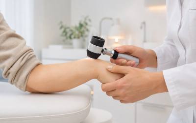
Dermatology is the branch of medicine concerned with the…
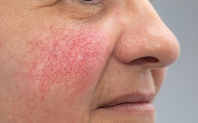
Rosacea is a chronic inflammatory skin condition that primarily affects…
Eczema is a general term for inflammatory skin conditions that…
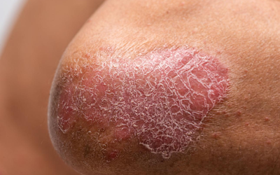
Psoriasis is a chronic, immune-mediated inflammatory condition that causes red,…
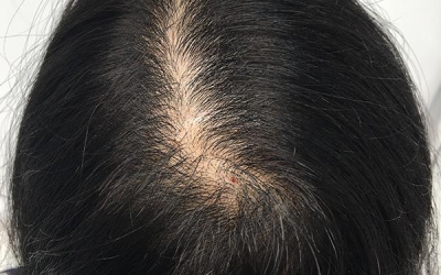
Hair loss is a common dermatologic concern that can affect…
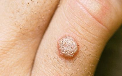
Warts are generally benign, contagious skin growths caused by certain…
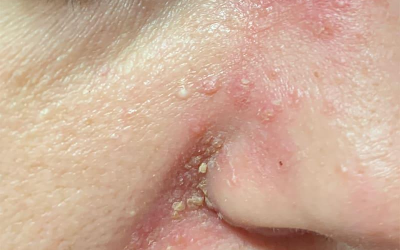
Seborrheic dermatitis is a chronic inflammatory skin condition that causes…
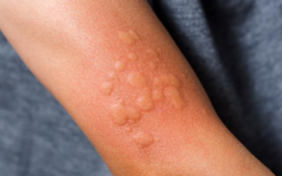
Urticaria, commonly known as hives, is a skin condition that…
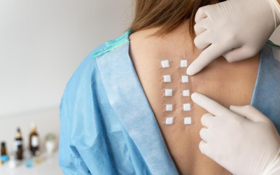
Allergic contact dermatitis is an inflammatory skin condition caused…
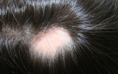
Alopecia areata is a condition in which the immune system…
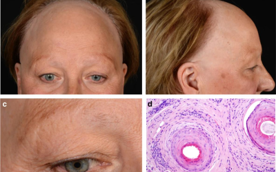
Eyebrow hair loss, medically known as eyebrow alopecia or madarosis,…
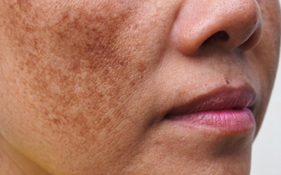
Melasma is a chronic pigmentation condition that causes brown or…
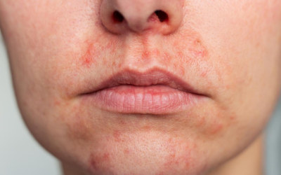
Perioral dermatitis is an inflammatory skin condition that causes small…
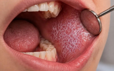
Lichen planus is a chronic, immune-related inflammatory condition that affects…

The effects of a product applied to the skin depend…

Human papillomavirus (HPV) is a common group of viruses…
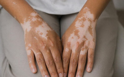
Vitiligo is a chronic skin condition characterized by white…

Acne is a chronic inflammatory skin condition that affects hair…